Home
Conference
Life Sciences sessions
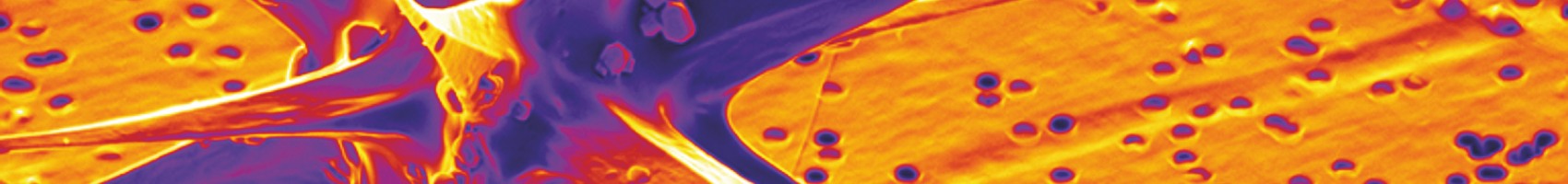
Frontiers in BioImaging: Correlative Light Electron Microscopy (CLEM)
Frontiers in BioImaging: Correlative Light Electron Microscopy (CLEM)
Session organisers: Dr Paul Verkade and Dr Raffaella Carzaniga
Tuesday 1st July from 14.15 - 16.15
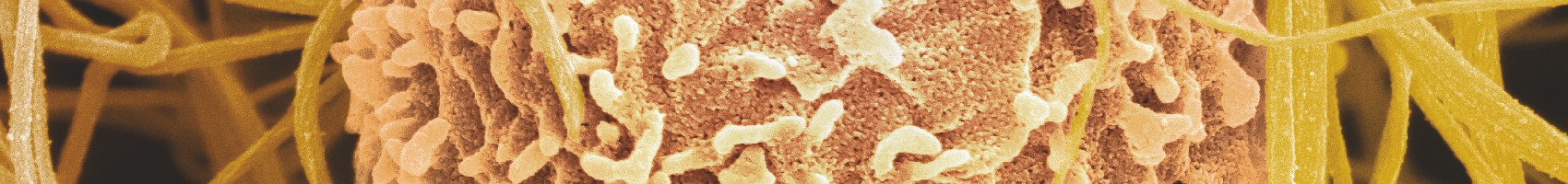
Correlative Microscopy aims to combine two (or more) techniques in one single experiment. At least one of the techniques in such an experiment will be based on Microscopy. Correlative Microscopy techniques have certain strengths over the application of the respective techniques separately. Probably the best-established Correlative Microscopy technique is the combination of light and transmission electron microscopy (CLEM). In this session we will be highlighting the latest developments in the field of CLEM, ranging from probes, sample processing, integrated techniques, and analysis. Other areas of Correlative Microscopy will be highlighted in separate sessions.
Invited Speaker:
Professor Pascal Fua
"Modeling Brain Circuitry over a Wide Range of Scales"
Dr Cristina Risco
"Metallothioneins for CLEM"